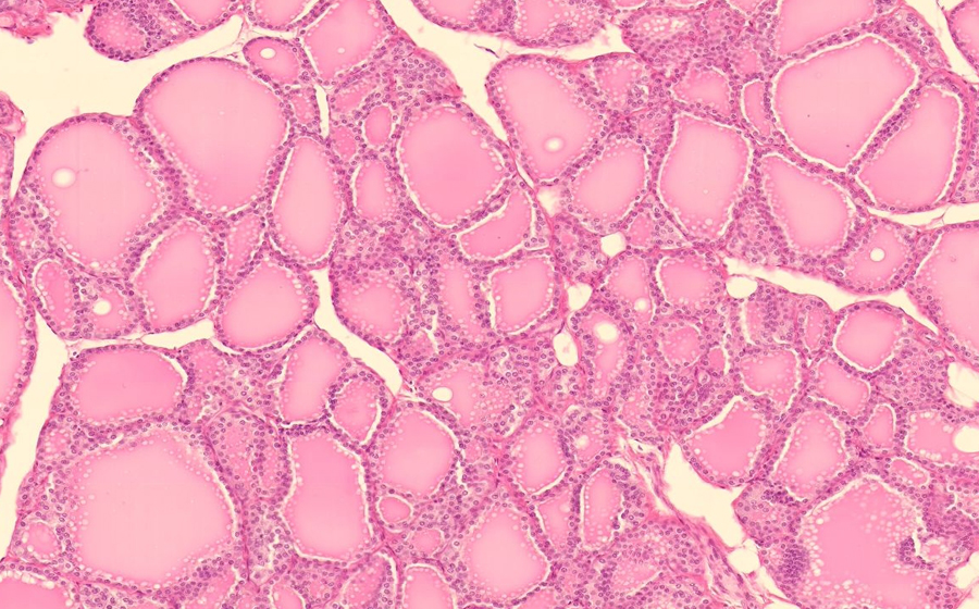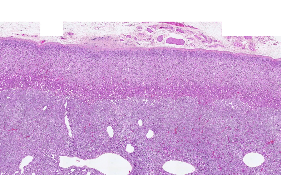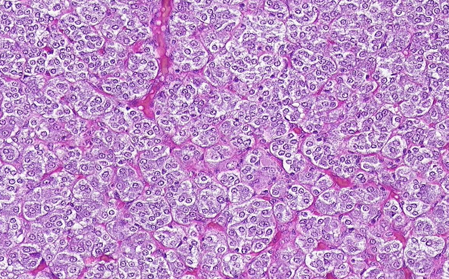
Image 1 (virtual): The pancreas was grossly unremarkable. A representative section is seen in the submitted slide.
At low power, the lobular structure of the pancreas can be well appreciated, created by loose connective that surrounds the surface of the pancreas, dissects into the parenchyma, and effectively subdivides it. Darker, eosinophilic cells predominate and compose the exocrine pancreas. Scattered among the exocrine pancreas are paler, eosinophilic, circular structures.
Question: What are the circular structures?
Question: What adjective can be assigned to histologic structures or material that have a somewhat uniform, homogeneous, eosinophilic, and/or proteineous quality?
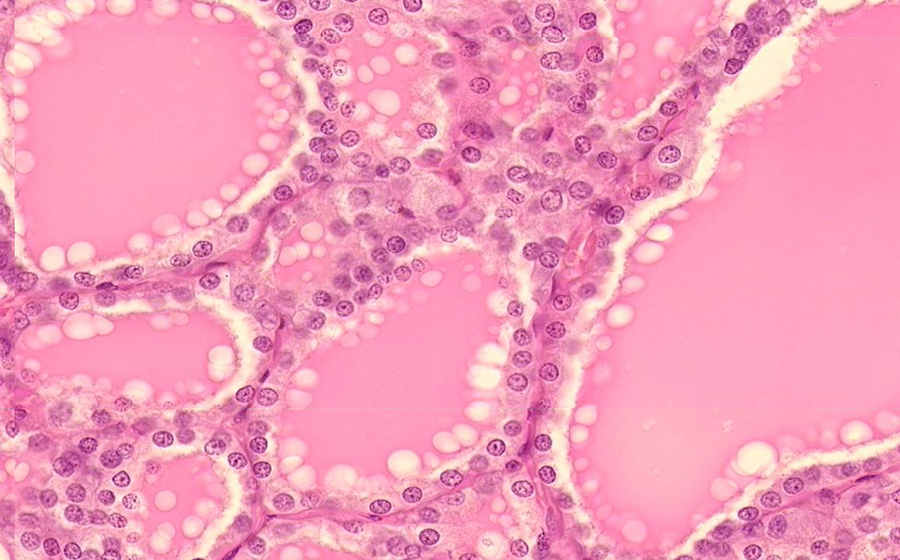
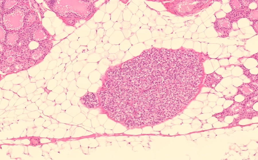
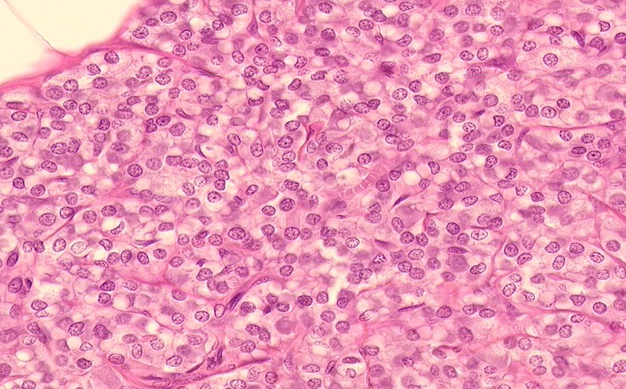
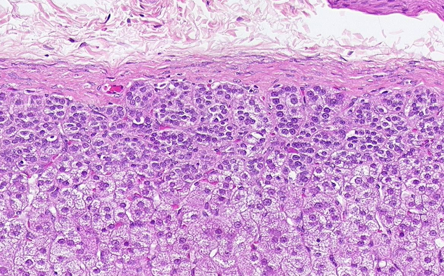
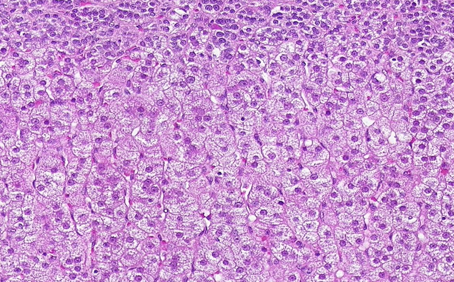
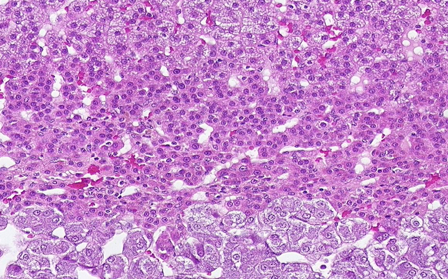
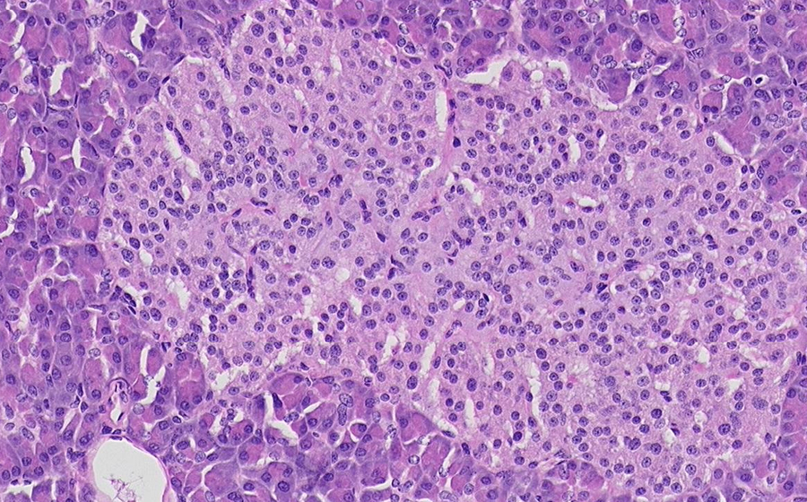
At high power, the structural unit of the exocrine pancreas, the acinus, is well visualized. A small cluster of acinar cells secrete their digestive enzymes into an intercalated duct, which eventually connects to a larger intralobular duct, the latter of which can be seen scattered throughout the exocrine parenchyma. Now identify one of the eosinophilic, circular islet of Langerhans, the architectural unit of the endocrine pancreas. Compared to their normal histology, these islets appear hyalinized and infiltrated by homogenous pink material. This material is composed of extracellular protein that is abnormally folded and deposited in various tissues in various disease states - the process of amyloidosis. Amyloid protein appears as small nodules that displace the islet cells into a ring-like arrangement around the deposits. Note the lack of inflammation or any other tissue reaction in response to the amyloid protein.
Question: What are the staining/microscopy methods for identifying amyloid?