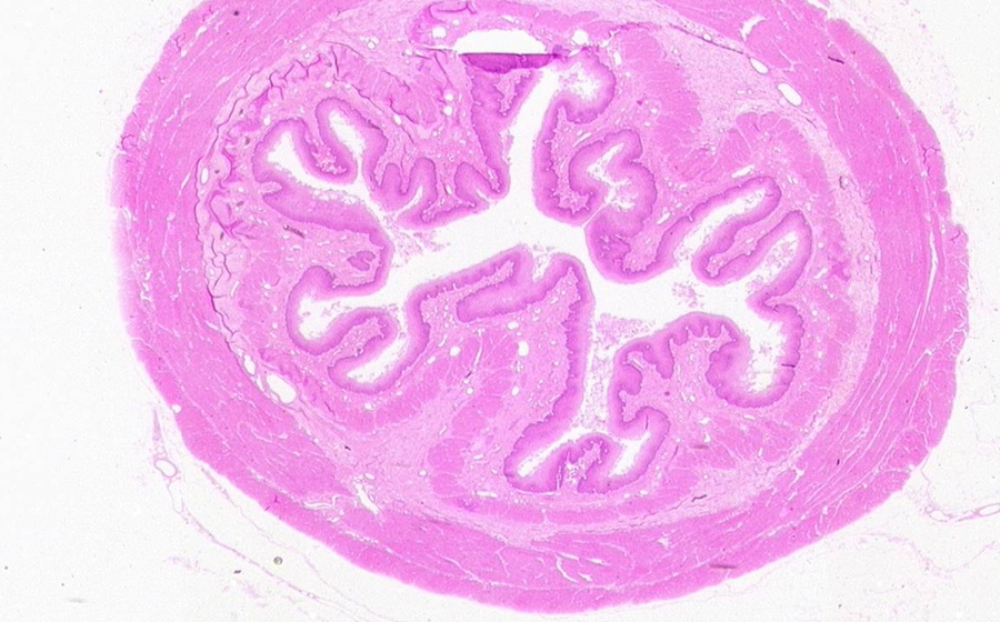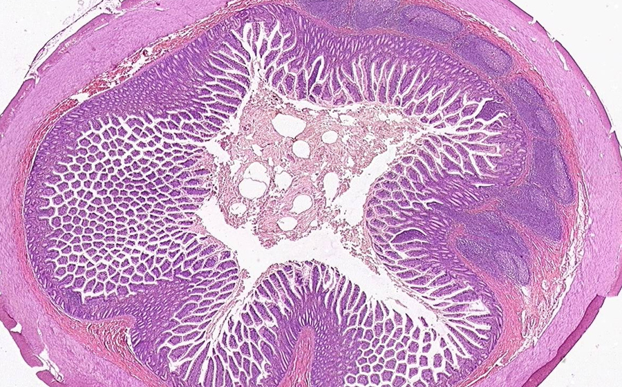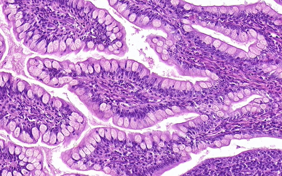
Image 1 (virtual): The histology of an abscess is nicely displayed in this slide, from a periappendiceal abscess. The abscess may have arisen in association with acute appendicitis, or from any type of acute (typically bacterial) intrapelvic process that just happens to involve the appendix or periappendiceal soft tissue. Low power evaluation reveals marked cellularity both in the portion of appendix (upper left), but also in the periappendiceal (mesenteric) adipose tissue. Congested vessels are also particularly prominent around the appendix. In the upper right portion of the abscess, an eosinophilic, hemorrhagic region is surrounded by relative basophilia.
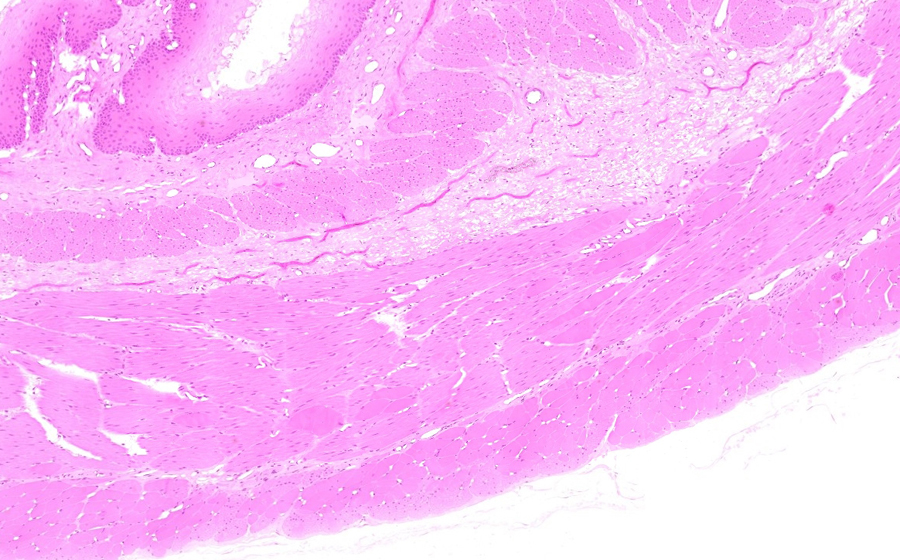
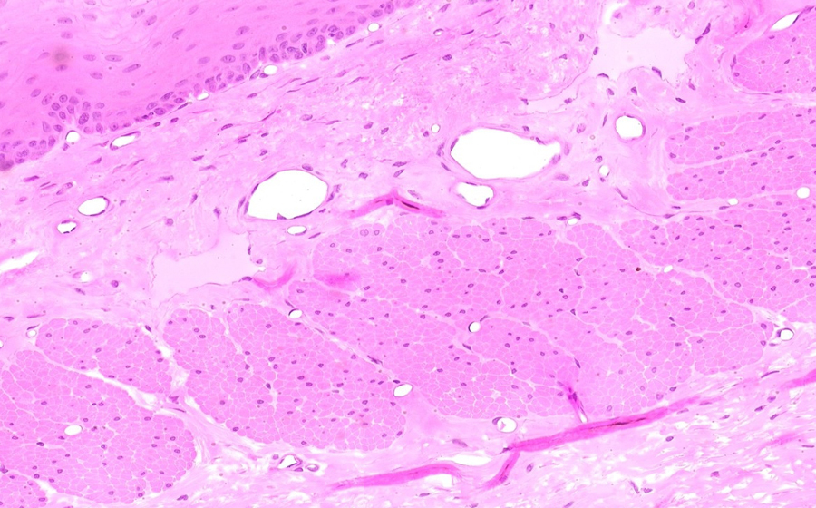
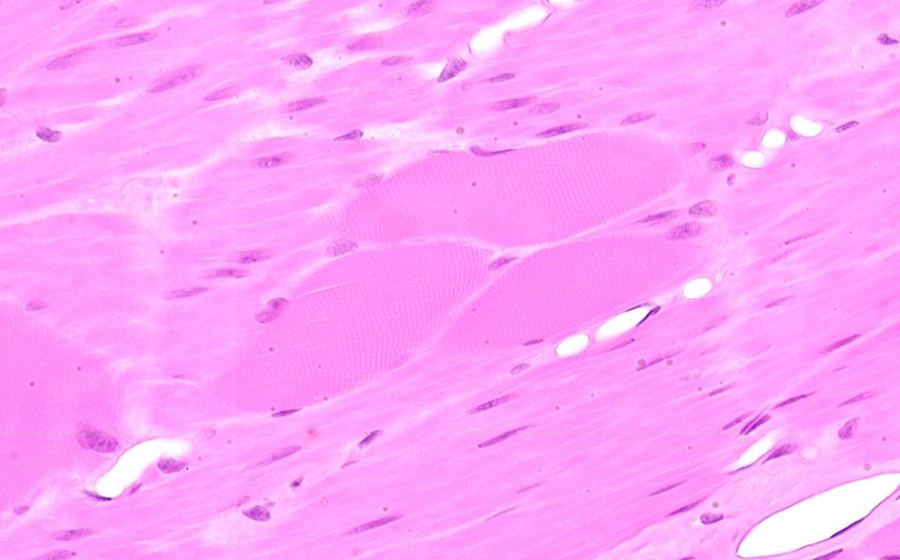
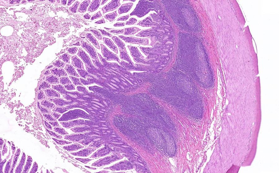
Higher power review of the upper right abscess shows a necrotic center consisting of abundant neutrophils, eosinophilic proteineous fluid, cell fragments and cell debris, and red blood cells. The basophilia seen at low power consists of a peripheral, partial rim of mononuclear cells, particularly lymphocytes. A single germinal center (lymphoid hyperplasia) is seen at the periphery of the large abscess. Also note there is a smaller necrotic focus in the (almost) direct center of the slide. The upper right portion of abscess may be large enough to appreciate grossly, but the center necrotic focus is likely only visible histologically (a 'microabscess').
The word abscess is used both to refer to a structure (as above), but also to a process. The process of abscess formation can be seen across the whole slide - i.e., the entirety of the inflammation-necrosis that is involving the appendix and periappendiceal adipose tissue.
Question: Describe the likely pathogenesis of this abscess development in our patient.