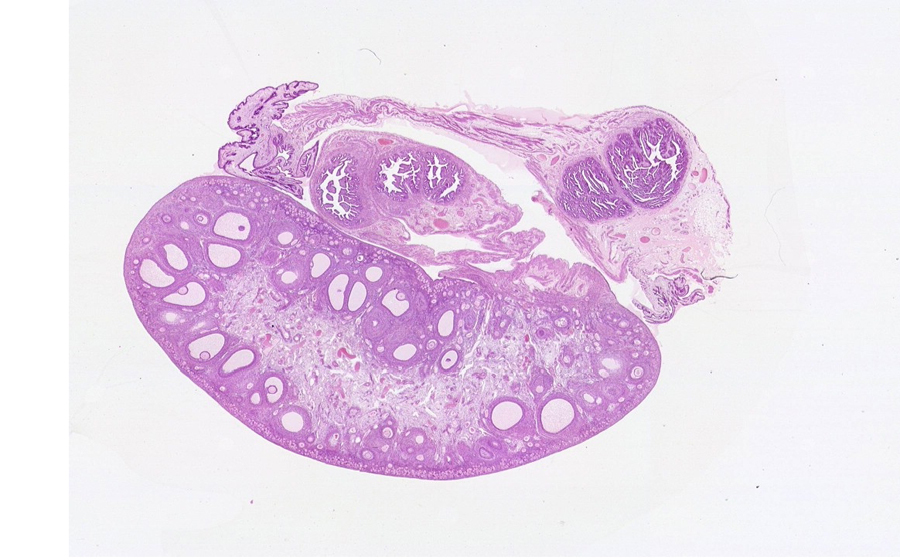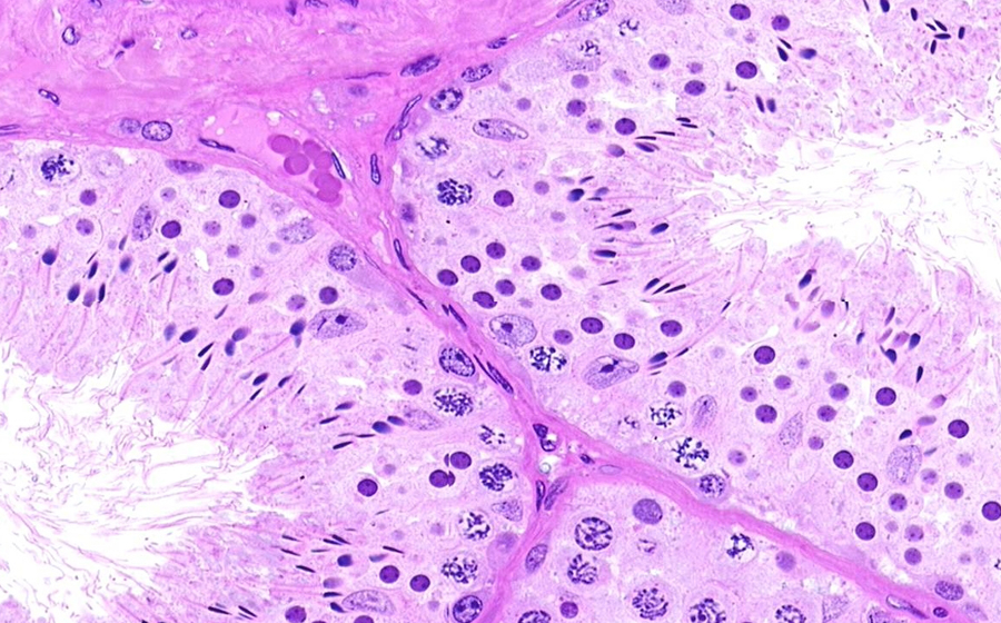
Image 1 (virtual): Grossly, one testicle was smaller than the other (right testicle: 20 grams; left testicle: 14 grams). A histologic section is procured through the patient's left testis.
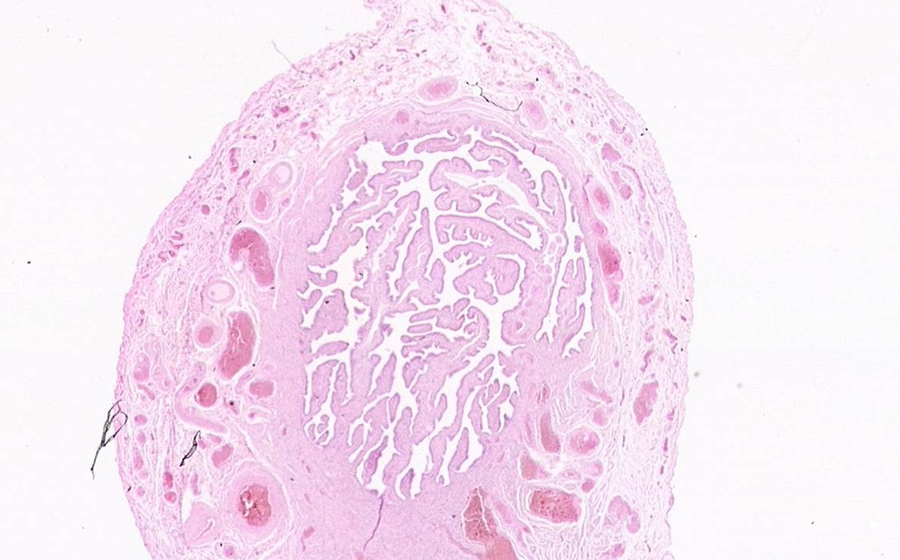
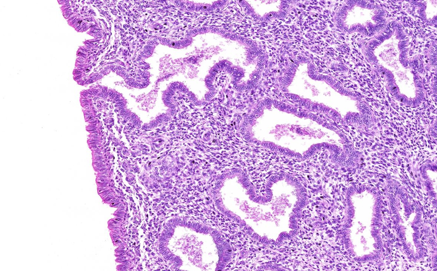
From low power, testicular parenchyma is seen with tunica albuginea on the far right of the slide. The histologic changes impart a hyaline appearance from low power; typically, the testes demonstrate more basophilia, due to the usual cellular abundance of intratubular spermatogenesis and its many cellular stages.
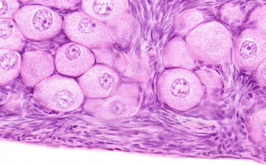
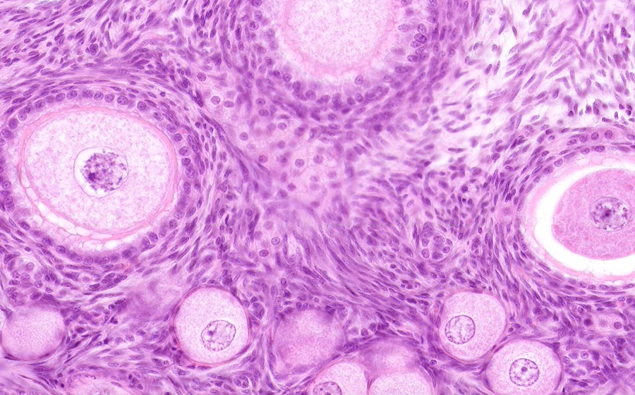
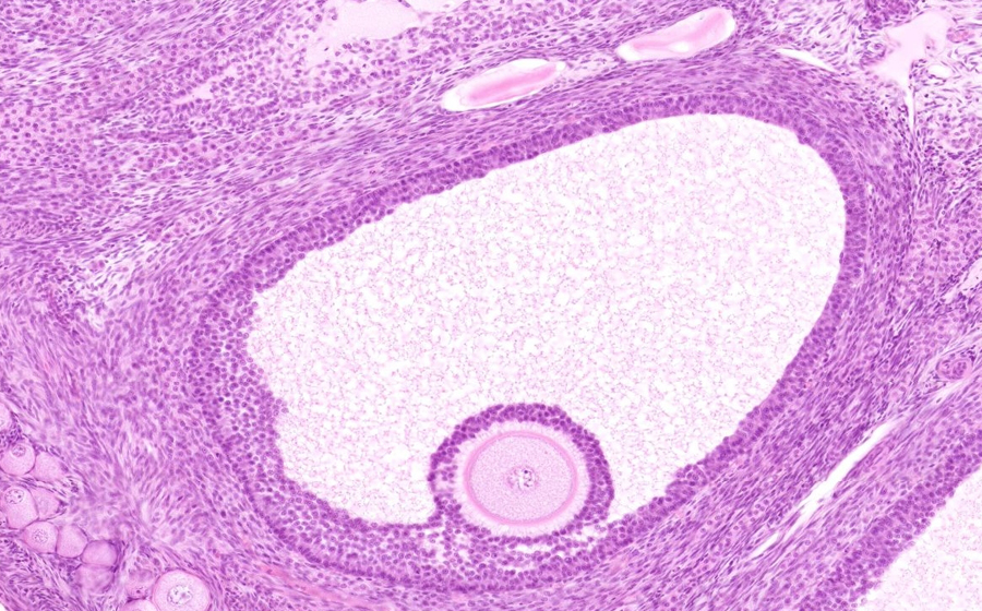
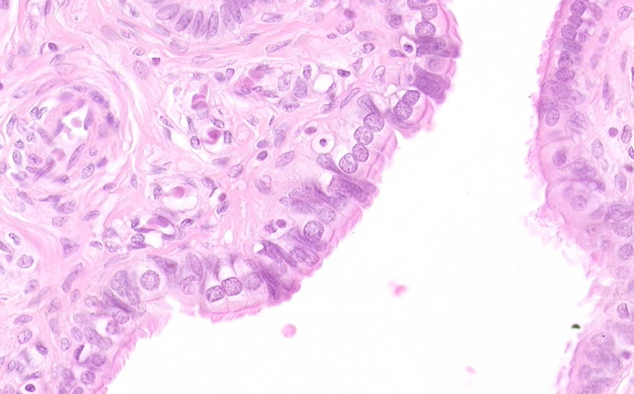
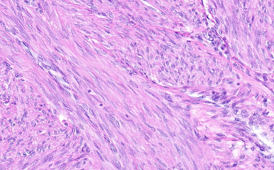
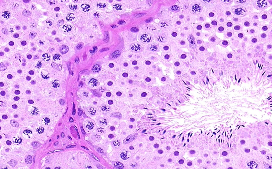
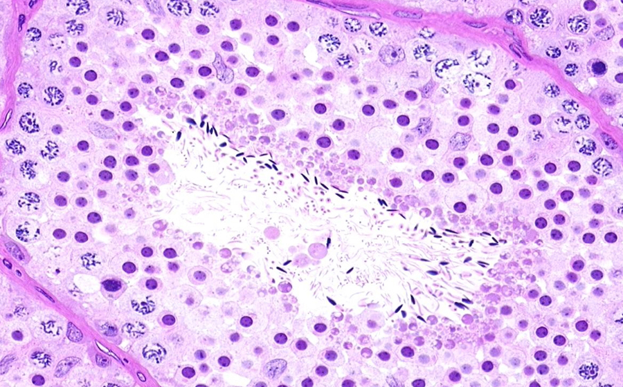
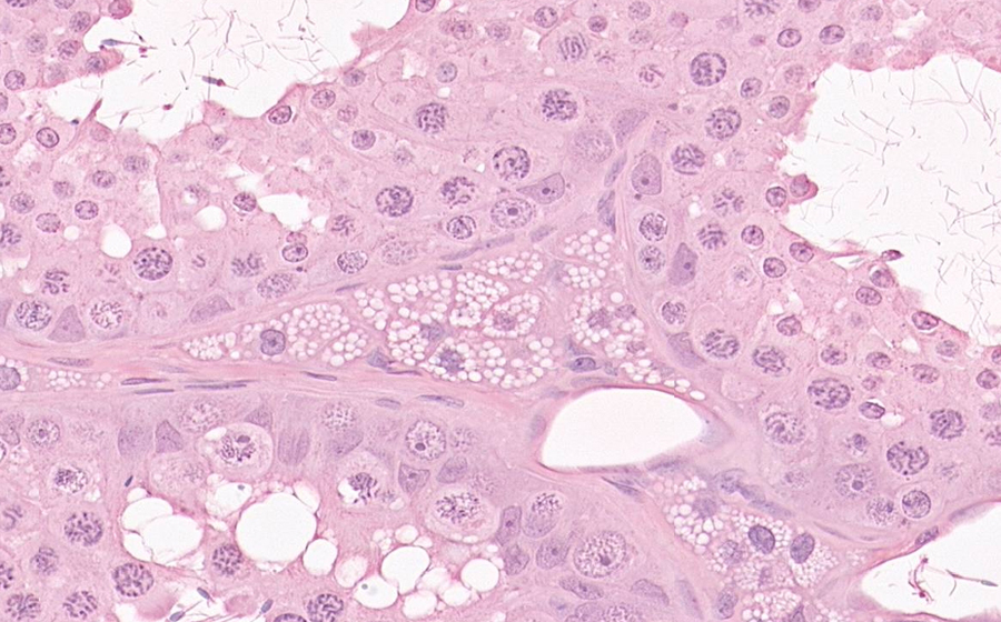
From higher power, the spectrum of changes associated with testicular atrophy can be appreciated. Particularly in the lower right of the slide, some tubules do show spermatogenesis. However, many tubules are completely hyalinized, characterized by collagen and fibroblast-like cellular replacement. Still others are intermediately affected with collagen and spermatogenic cells mixed within the same tubule. Also note that eosinophilia is imparted by Leydig cell hyperplasia diffusely occurring throughout the tissue. Leydig cell clusters expand the interstitium, but do not distort, compress, or destroy the seminiferous tubules.
Question: What could be some etiologies of atrophy in our patient?