Topic 8: Synapses
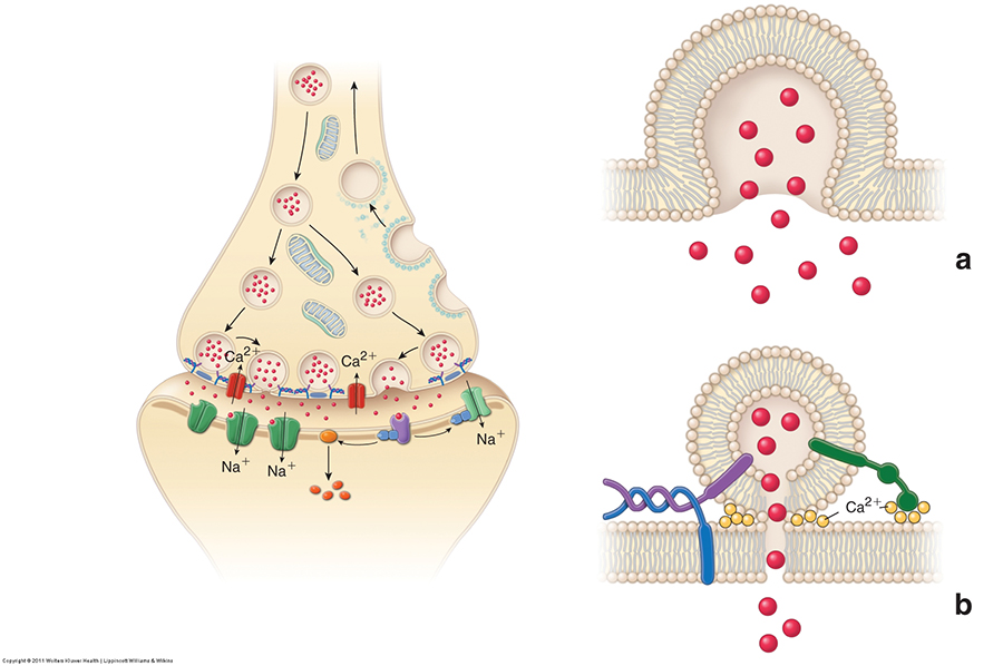
Chemical axodendritic synapse
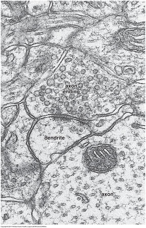
EM - Nerve process in cerebral cortex
Ross & Pawlina, Histology Text & Atlas - Figures 12.7 & 12.8
Structure of CNS synapses
EM image of components of axo-dendritic synapses
|
|
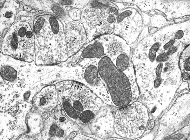
Axo-dendritic synapses, EM
Figure legend:
Occupying the center of the field is a large spine or protrusion from the perikaryon of a neuron, and surrounding this structure is a variety of axon terminals. Two of the terminals (At1 and At2) contain small, spherical synaptic vesicles, while the spherical vesicles in two other teminals (At3 and At4) are somewhat larger. The other axon terminals (At4 to At9) contain some elongate synaptic vesicles. The synaptic junctions in which these various axon terminals participate all seem to be symmetrical. The junction formed by At5 is noteworthy because of the regular array of presynaptic densities (arrows). At the other synaptic junctions, only one or two of these densities (arrows) are apparent. Ventral cochlear nucleus of adult rat X44,000.
Click on image to magnify |
|
From: Peters et al., The Fine Structure of the Nervous System, W.B. Saunders Co., Philadelphia, 1976
End of Topic 8